Trigeminal Neuralgia
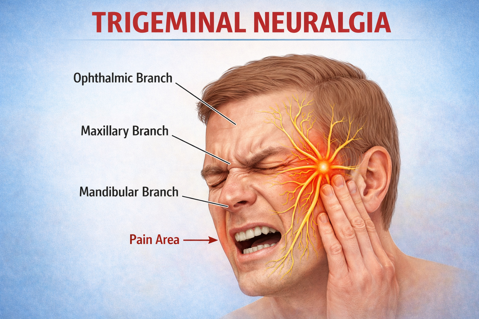
Trigeminal Neuralgia (TN) is a severe chronic pain condition affecting the fifth cranial nerve (trigeminal nerve), which provides sensation to the face. Often described as one of the most agonizing painful conditions in medicine, it causes sudden, shock-like, or burning facial pain triggered by mild tactile stimulation such as chewing, speaking, washing the face, or brushing teeth.
Dr. Sanjeev Srivastava, Director of Neurosurgery at CK Birla Hospital, Gurgaon, treats Trigeminal Neuralgia through advanced microsurgical and minimally invasive techniques aimed at relieving neural compression and restoring normal nerve function.
Causes & Pathophysiology
Vascular Compression (Primary TN): In the vast majority of cases, an adjacent blood vessel (most commonly the superior cerebellar artery) presses against the root entry zone of the trigeminal nerve near the brainstem, causing demyelination and hyperexcitability.
Secondary Causes: Structural lesions such as cerebellopontine angle (CPA) meningiomas, epidermoid cysts, or multiple sclerosis plaques affecting the trigeminal nerve pathway.
Treatment Options Offered by Dr. Sanjeev Srivastava
| Treatment Modality | Procedure Type | Mechanism / Indication | Primary Advantage |
| Medical Management | Pharmacotherapy | First-line treatment using sodium channel blockers (Carbamazepine, Oxcarbazepine) or Gabapentinoids. | Non-invasive initial pain control. |
| Microvascular Decompression (MVD) | Open Skull Base Surgery | Retrosigmoid craniotomy to isolate the offending vessel from the nerve using a Teflon felt sponge insert. | Highest long-term success rate; preserves facial sensatio. |
| Percutaneous Rhizotomy | Minimally Invasive | Needle inserted through the cheek into the Foramen Ovale using balloon compression, radiofrequency, or glycerol ablation. | Option for elderly or high-risk patients unsuitable for open surgery. |
| Stereotactic Radiosurgery (SRS) | Non-Invasive Radiation | High-dose targeted radiation (Gamma Knife/CyberKnife) focused on the trigeminal nerve root. | No surgical incision required; gradual pain relief over weeks. |
Clinical Evaluation & Surgical Workflow
Diagnostic Mapping & High-Resolution Imaging
3T MRI with thin-slice heavy T2 sequences (FIESTA/CISS) to visualize exact arterial/venous contact with the trigeminal nerve
↓
Multidisciplinary Case Assessment
Evaluation of pain response to first-line anticonvulsant medications and patient surgical candidacy
↓
Surgical / Minimally Invasive Intervention
Microvascular Decompression (MVD) via retrosigmoid keyhole approach or percutaneous/radiosurgical neuro-ablation
↓
Post-Operative Recovery & Pain Monitoring
Immediate post-surgical pain resolution assessment, gradual tapering of oral pain medications, and long-term follow-up