Aneurysms
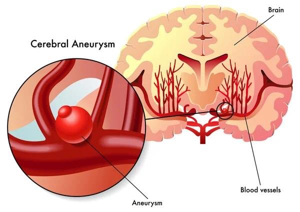
An aneurysm is an abnormal bulging or ballooning of a weak area in the wall of a blood vessel, usually an artery.
Dr. Sanjeev Srivastava, Director of Neurosciences, specializes in the comprehensive management of cerebral aneurysms. His practice encompasses both micro-neurosurgical clipping and minimally invasive endovascular interventions.
Core Types of Cerebral Aneurysms Treated
Saccular (Berry) Aneurysms: The most common form, appearing as a rounded sac protruding from a cerebral arterial bifurcation (most frequently along the Circle of Willis).
Fusiform Aneurysms: Circumferential widening and elongation of the arterial wall without a distinct neck, often requiring complex bypass or flow-diversion strategies.
Mycotic (Infectious) Aneurysms: Rare vascular bulges caused by infectious emboli damaging the arterial wall, requiring targeted antimicrobial therapy combined with surgical or endovascular management.
Unruptured vs. Ruptured Aneurysms: Unruptured aneurysms are often managed electively based on size, location, and rupture risk, whereas ruptured aneurysms cause sudden subarachnoid hemorrhage (SAH) and require emergency intervention to prevent re-bleeding.
Treatment Modalities
| Modality | Procedural Approach | Key Indications / Advantages |
| Microsurgical Clipping | Open craniotomy; a micro-titanium clip is placed across the aneurysm neck under direct microscopic view. | Complete anatomical isolation, low long-term recurrence rates, preferred for wide-necked or middle cerebral artery (MCA) aneurysms. |
| Endovascular Coiling | Percutaneous arterial catheterization; micro-coils are packed inside the aneurysm sac. | Minimally invasive (no craniotomy), faster recovery, ideal for deep-seated basilar or posterior circulation aneurysms. |
| Flow-Diverting Stents | Endovascular placement of a dense mesh stent across the parent artery. | Redirects blood flow past giant or fusiform aneurysms, promoting gradual thrombosis and arterial remodeling. |
Clinical Management Workflow for Cerebral Aneurysms
Diagnostic Mapping & Risk Assessment
High-resolution CTA, MRA, or 3D Digital Subtraction Angiography (DSA) to evaluate aneurysm size, shape, neck geometry, and rupture risk
↓
Multidisciplinary Case Selection
Treatment selection (clipping vs. coiling vs. flow diversion) based on patient age, medical comorbidities, and vascular anatomy
↓
Surgical or Endovascular Repair
Microsurgical craniotomy for clip application or neuro-interventional access via femoral/radial arteries for coil delivery/stenting
↓
Post-Operative Neuro-ICU Management
Hemodynamic monitoring, vasospasm prevention (hypervolemic/nimodipine protocol), and follow-up angiography to confirm complete exclusion