
Hydrocephalus
Hydrocephalus is a condition characterized by an abnormal accumulation of cerebrospinal fluid (CSF) within the brain’s ventricles. This accumulation increases intracranial pressure (ICP), causing ventricular expansion and progressive compression of surrounding brain tissue.
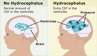
Etiology and Classification
Hydrocephalus can develop across all age groups—from congenital infant cases to adult Normal Pressure Hydrocephalus (NPH)—and is classified into three core types based on underlying mechanism:
Communicating (Non-Obstructive) Hydrocephalus: Occurs when CSF flows freely through the ventricular system, but absorption into the venous circulation via arachnoid granulations is impaired (e.g., secondary to subarachnoid hemorrhage, meningitis, or head trauma).
Non-Communicating (Obstructive) Hydrocephalus: Occurs when physical structural blockade prevents CSF from flowing between ventricles (e.g., aqueductal stenosis, colloid cysts, or posterior fossa brain tumors).
Normal Pressure Hydrocephalus (NPH): Primarily affects older adults, presenting as ventricular enlargement with intermittently normal or mildly elevated CSF pressure. It classically presents with the clinical triad: gait instability, urinary incontinence, and cognitive decline.
Surgical Management Options
| Treatment Modality | Indications / Mechanism | Key Operational Features |
| Ventriculoperitoneal (VP) Shunt | Communicating and non-communicating hydrocephalus | Placement of a silicone catheter system with a pressure-programmable valve that diverts excess CSF from the lateral ventricle to the peritoneal cavity. |
| Endoscopic Third Ventriculostomy (ETV) | Obstructive hydrocephalus (e.g., aqueductal stenosis) | Endoscopic keyhole procedure creating a small perforation in the floor of the third ventricle to establish an internal CSF bypass, avoiding permanent hardware implantation. |
| Ventriculoatrial (VA) / Ventriculopleural Shunt | Refractory hydrocephalus where peritoneal absorption is impaired | Alternative shunt trajectories diverting CSF into the right atrium of the heart or pleural cavity. |
Hydrocephalus Surgical & Clinical Management Workflow
Diagnostic Evaluation & Volumetric Imaging
High-resolution non-contrast CT, epilepsy/hydrocephalus protocol MRI, or lumbar tap test (in suspected NPH) to confirm ventricular enlargement and assess CSF dynamics
↓
Surgical Approach Selection
Multidisciplinary determination choosing between internal endoscopic bypass (ETV) for obstructive etiology or programmable shunt placement (VP Shunt)
↓
Micro-Neurosurgical / Endoscopic Procedure
Stereotactic image-guided ventricular catheter placement or endoscopic fenestration of the third ventricle floor under direct high-definition visualization
↓
Post-Operative Valve Titration & Long-Term Surveillance
Non-invasive external magnetic valve adjustment, serial neurological assessment, and long-term surveillance imaging to ensure optimal CSF drainage